| What is arthroscopy? |
| Arthroscopy is a surgical procedure by which the internal structure of a joint is examined for diagnosis and/or treatment using a tube-like viewing instrument called an arthroscope. Arthroscopy was popularized in the 1960s and is now commonplace throughout the world. Typically, it is performed by orthopedic surgeons in a day care setting.
The technique of arthroscopy involves inserting the arthroscope, a small tube that contains optical fibers and lenses, through tiny incisions in the skin into the joint to be examined. The arthroscope is connected to a video camera and the interior of the joint is seen on a television monitor. The size of the arthroscope varies with the size of the joint being examined. For example, the knee is examined with an arthroscope that is approximately 4 or 5 millimeters in diameter. There are arthroscopes as small as 0.5 millimeters in diameter to examine small joints such as the wrist. If procedures are performed in addition to examining the joint with the arthroscope, this is called arthroscopic surgery. There are a number of procedures that are done in this fashion. If a procedure can be done arthroscopically instead of by traditional surgical techniques, it usually causes less tissue trauma, results in less pain, and may promote a quicker recovery. |
 |
| For what diseases or conditions is arthroscopy considered? |
| Arthroscopy can be helpful in the diagnosis and treatment of many non-inflammatory, inflammatory, and infectious types of arthritis as well as various injuries within the joint.
Non-inflammatory degenerative arthritis, or osteoarthritis, can be seen using the arthroscope as frayed and irregular cartilage. Recently, for isolated cartilage wear in younger patients, repair of crevasses in the cartilage, using a “paste” of a patient’s own cartilage cells harvested and grown in the laboratory, has been performed using an arthroscope. In inflammatory arthritis, such as rheumatoid arthritis, some patients with isolated chronic joint swelling can sometimes benefit by arthroscopic removal of the inflamed joint tissue (synovectomy). The tissue lining the joint (synovium) can be biopsied and examined under a microscope to determine the cause of the inflammation and discover infections, such as tuberculosis. Arthroscopy can provide more information in situations which cannot be diagnosed by simply aspirating (withdrawing fluid with a needle) and analyzing the joint fluid. Common joint injuries for which arthroscopy is considered include cartilage tears (meniscus tears), ligament strains and tears, and cartilage deterioration underneath the kneecap (patella). In knee joint ACL reconstruction can be done arthroscopically. In younger patients Meniscal repairs can be done. Surgery for Recurrent Dislocation of Patella is also performed arthroscopically. In shoulder joint , Sub-Acromial Decompression, Stabilisation for Recurrent Dislocation of Shoulder / Shoulder Instability is also possible. Arthroscopy is commonly used in the evaluation of knees and shoulders but can also be used to examine and treat conditions of the wrist, ankles, and elbows. Finally, loose tissues, such as chips of bone or cartilage, or foreign objects, such as plant thorns, that become lodged within the joint can be removed with arthroscopy. |
| What is done in preparation for arthroscopy? |
| Arthroscopy is essentially a bloodless procedure and generally has few complications. The underlying health of the patient is considered when determining who is a candidate for arthroscopy. Most importantly, the patient should be able tolerate the anesthetic that is used during the procedure. A person’s heart and lung function should be adequate. If there are existing problems such as heart failure or emphysema, these should be optimized as possible prior to surgery. Patients who are on anticoagulants (blood thinners) should have these medications carefully adjusted prior to surgery. Other medical problems should also be controlled prior to surgery, such as diabetes and high blood pressure.
Preoperative evaluation of a patient’s health will generally include a physical examination, blood tests, and a urinalysis. Patients who have a history of heart or lung problems and generally anyone over the age of 50 will usually be asked to obtain an electrocardiogram (ECG) and a chest X-ray. Any sign of ongoing infection in the body usually postpones arthroscopy, unless it is being done for possible infection of the joint in question. |
| How is arthroscopy performed? |
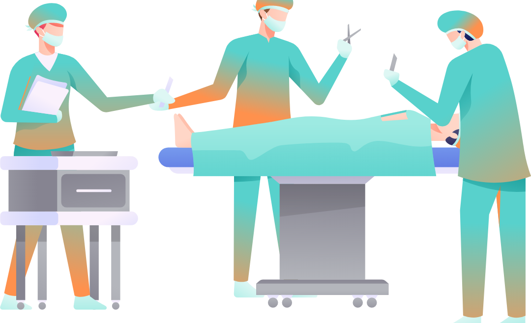
| Arthroscopy is most often performed as an outpatient procedure. The patient will check into the facility where the procedure is being performed and an intravenous line (IV) established in order to administer fluids and medication. The type of anesthesia used varies depending on the joint being examined and the medical health of the patient. Arthroscopy can be performed under a general anesthetic, a spinal or epidural anesthetic, a regional block (where only the extremity being examined is numbed), or even a local anesthetic. After adequate anesthesia is achieved, the procedure can begin. An incision is made on the side of the joint to be examined and the arthroscope is inserted into the incision. Other instruments are sometimes placed in another incision to help manoeuvre certain structures into the view of the arthroscope. In arthroscopic surgery, additional instruments for surgical repairs are inserted into the joint through the arthroscope. These instruments can be used to cut, remove, and sew damaged tissues. Once the procedure is completed, the arthroscope in removed and the incisions are sutured (sewn) closed. A sterile dressing is placed over the incision and a brace wrap may be placed around the joint. |
| How does the patient recover after arthroscopy? |
| Immediately after arthroscopic surgery, patients may be sleepy, especially if a general anesthetic has been used. Medications are administered to control pain if needed. If a local anesthetic has been used, there may be no pain at all immediately after the procedure. If a spinal or regional anesthetic has been used, there can be numbness and weakness of the extremity that gradually resolves before the patient is sent home.
The surgical incisions from arthroscopy are small. They usually consist of two mm (1/4 inch) incisions on either side of the joint, which are bandaged after surgery. The bandage may absorb some of the tissue drainage from these wound sites. The bandage should only be removed under the guidance of the treating surgeon or nurse. It should otherwise be kept as dry as possible during the first few days after surgery. Patients should notify their physician’s office immediately if they develop unusual joint pain, fever swelling, redness or warmth, or if they injure the involved joint. For several days after arthroscopy, patients will generally be asked to rest and elevate the joint while applying ice packs to minimize pain and swelling. After surgery, an exercise program is gradually started that strengthens the muscles surrounding the joint and prevents scarring (contracture) of surrounding soft tissues. The goal is to recover stability and strength of the joint rapidly and safely, while preventing the build-up of scar tissue. This program is an essential part of the recovery process for an optimal outcome of this procedure. Over the years, higher quality fiber-optic equipment has allowed the development of miniature arthroscopes. This has allowed the examination of smaller joints with arthroscopy. Arthroscopy has become an integral tool for orthopedic surgery and its role will continue to expand as further improvement in arthroscopes and arthroscopic instruments continues. |
| Arthroscopy At A Glance |
|

